Comparison of Morphological Analysis of RBC through Peripheral Smear and Automated Method-Juniper Publishers
JUNIPER PUBLISHERS-OPEN ACCESS GLOBAL JOURNAL OF PHARMACY & PHARMACEUTICAL SCIENCES
Aim: To compare the morphology of red blood cells by peripheral smear and automated method.
Objective: Comparison of morphology of red blood cells by using peripheral smear and automated method.
Background: Counting of red blood cells (RBC)
in blood cell images is very important to detect as well as to follow
the process of treatment of many diseases like anaemia, leukaemia etc.
However, locating, identifying and counting of -red blood cells manually
are tedious and timeconsuming that could be simplified by means of
automatic analysis.
Reason: To know which is the most effective method of analysing morphology of RBC.
Keywords: Red blood cells; Observers; Automated method
Introduction
Red blood cells (RBCs), also known as erythrocytes,
are the most common type of blood cells in our body and it is the
principal means of delivering oxygen to the body tissues.The red blood
cells are typically biconcave disks: flat and depressed in the centre,
with a dumbbell-shaped cross section, and a rim on the edge with the
torus shapeddisk [1].
Red blood cells in mammals are unique amongst vertebrates as they do
not have nuclei when mature.Anaemia is the lack of red blood cells and/
or haemoglobin. This results in a reduced ability of blood to transfer
oxygen to the tissues [2].
Normal, mature RBCs are biconcave, disc-shaped, a nuclear cells
measuring approximately 7-8 microns in diameter on a peripheral blood
smear with an internal volume (MCV) of 80-100 femto liters (fL). The
term used to describe RBCs of normal size is "normocytic". Anaemia is
classified by the size of the red blood cells which is either done
automatically or on microscopic examination of a peripheral blood smear.
The size is reflected in the mean corpuscular volume (MCV). If the
cells are smaller than normal (under 80fl), the anemia is said to be
microcytic; if they are normal size (80- 100fl), normocytic; and if they
are larger than normal (over 100fl), the anaemia is classified as
microcytic [3].
MCV measures only average cell volume. The MCV can be normal while the
individual red cells of the population vary wildly in volume from one to
the next. Such an abnormal variation in cell volume is called
anisocytosis [1]. The degree of anisocytosis in a sample of blood is known as the red cell distribution width (RDW).
Materials and Methods
The study was done using 50 blood smears of patients with different age groups.
Blood samples
All venous blood specimens were collected in tubes containing ethylenediaminetetraacetic acid (K3EDTA) and then were analysed
Automated method
After thorough mixing of each blood sample on an
automated mixer for 3-5min, a mean corpuscular value was obtained in
which value between 80-100 were considered normal.
Manual method
Thin air-dried blood smears made after thorough
mixing of each sample were stained manually with leishman’s stain and
examined under light microscopy with a X100 oil-immersion lens.
Results and Discussion
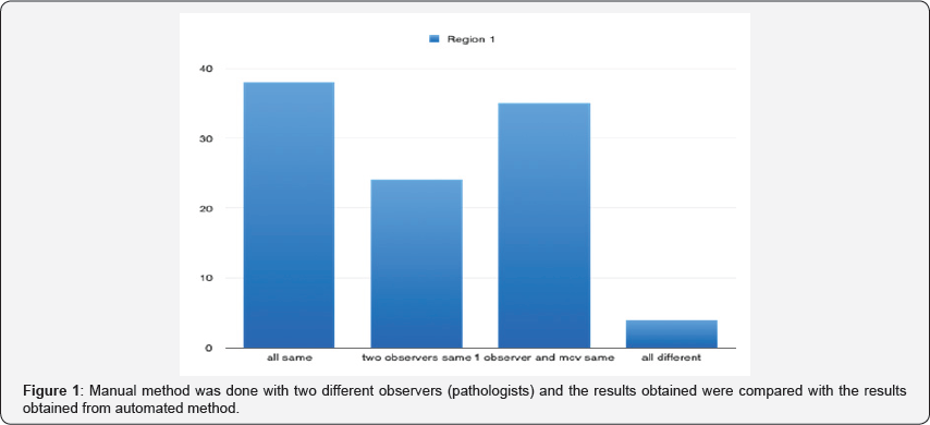
Manual method was done with two different observers
(pathologists) and the results obtained were compared with the results
obtained from automated method. The similarities among the results are
shown in the graph shown in Figure 1.
Out of 50, 38% of the results were same with both the observes and the
automated method. 24% of the results were same between 2 observers 17%
of the results were common between one observer and the automated value.
Only 4% of the results were different among all the observers and the
automated method. Among all 50 slides when examined by the observerl, 24
slides were normocytic, in case of observer 2, 39 were normocytic and
in automated method 26 were normocytic. Most cases of normocytic anemia
are caused by blood loss, suppressed production of RBCs, or hemolysis [2].
Macrocytic cells were seen in 9 slides by observer 1, only one slide
had macrocytic cells by observer 2 and 3 slides in automated method.
Macrocytic is usually seen to differentiate between megaloblastic and
nonmegaloblastic causes megaloblastosis is seen with and folate
deficiency, MDS and CDA, HIV infection, and rare inborn errors of
metabolism, while nonmegaloblastic causes include liver and thyroid
disease, alcohol, Down syndrome, aplastic anemia, and reticulocytosis.
Medications can be responsible for both megaloblastic and
non-megaloblastic anemia, while RBC agglutination may lead to spurious
macrocytosis [2].
Microcytic cells are seen in 17 slides by observer 1,10 slides had
microcytic cells by observer 2 and 20 slides were microcytic in
automated method.In classic cases, the morphological differentiation of
the three common microcytic anemias is straightforward [4].

Conclusion
The review of red blood cell morphology is the most
important step in the evaluation of a patient with anemia. It can be
very useful in evaluating microcytic, normocytic, and macrocytic anemias
and is especially helpful in the patients with hemolysis [5].
It can be concluded that for diagnostic purposes results obtained from
two different observers or results from both the observers and the
automated value can be considered.
For more Open Access Journals in Juniper Publishers please click on: https://juniperpublishers.com
For more articles in Global Journal of Pharmacy & Pharmaceutical Sciences please click on: https://juniperpublishers.com/gjpps/index.php
To know more about Juniper Publishers please click on: https://juniperpublishers.business.site/


Comments
Post a Comment